MIP Panos

eric....here are the panos....what do you think?
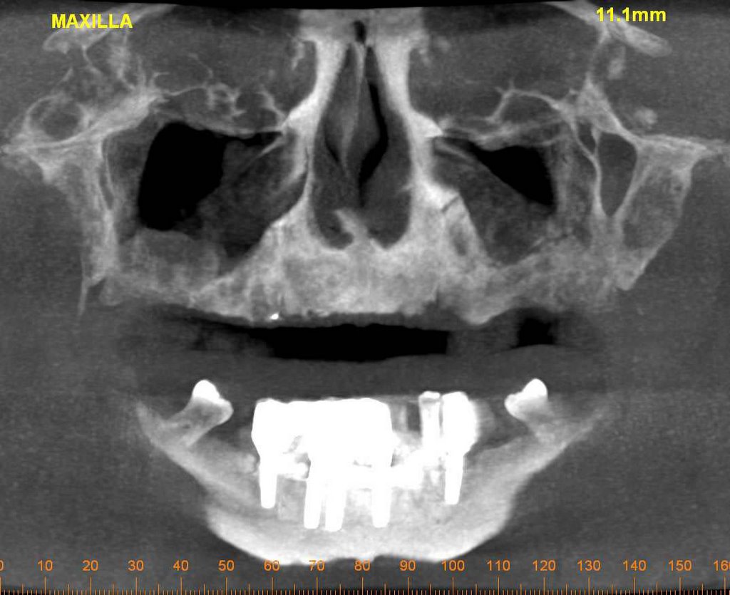
We recently did a MIP pano for maxillary implants sites. Patient had some fluid, mucoceles, or some pathology in sinus (which of course we see pretty often). In this case the patient had very little bone - literally a few mm in some places. When we did a 14mm MIP the fluid (or whatever) in the sinus appears as bone. So, the doc calls and says "what is this? why do I have bone in this pano and not in the other?" Our standard answer is always to use the cross sectional views as the gospel - not pano views. But it was odd. Any comments? What exactly are we seeing in the MIP view?
Also, for the pano view for maxillary implants - we are only mapping the maxilla (duh...). We used to map all the way back to the TMJ just to get the traditional "pano" look but it causes more problems because the mandible is so distorted - or we split the difference and the maxilla is not as accurate. Am I a little slow to figure this out?
posted by Marcelle at 12/12/2005 10:06:00 AM
![]()
![]()

4 Comments:
Marcelle and Vicki:
Would you kindly add the the MIP pan views to the blog so we can see exactly what you are questioning?
Often, I map out the mandible and then move the map to the maxilla and see how it looks. If necessary, I adjust it, mostly to center the cross sections in the window. I take most of my implant scans with the patient biting edge to edge on a cotton roll, which seems to put the 2 arches in pretty much the same focus.
Maximum Intensity Projection (MIP)
The MIP is an algorithm (a procedure consisting of a sequence of algebraic formulas and/or logical steps to calculate or determine a given task) that is used to extract high intensity structures from volumetric data. MIP assigns an intensity to each pixel that is the maximum of all intensities (for example-bone) encountered along a line through that pixel. You will not be able to see pixels behind the maximum intensity pixel because that intensity will be lower, thus not visible. Other anatomical structures and their projection path (depending on their intensity and size) might also interfere with your region of interest (ROI), which appears to have occurred in your posted example. The MIP does not give good depth perception because it is essentially a collapsed 3-D structure on a 2-D surface without any perspective. Your MIP image is a prime example, as it ‘appears’ the bone and sinus fluid (or whatever that is in the sinus) seems to be in the same plane and almost the same density, when in reality it is not.
A much thinner Slice Thickness (ST) in the oblique (panoramic) view would eliminate some of the interference in the MIP projection, however, the resultant view might not adequately demonstrate the ROI. As you indicated, for complete interpretation the best method to compensate for the loss of information or increased interference from other structures is to view the images in cross sectional and multiplanar reformatted (MPR) images.
When Imaging Sciences launches iCATVision, it will enable your referring dentist to easily view the anatomical structures to make a complete confident diagnosis without incurring any additions cost to unlock the study. It will take time and education for everyone to really comprehend i-CAT’s full capability. In the meantime, it is highly advisable that you let your dentist know that perhaps he/she should consider the option of having the entire scan read by an oral radiologist.
To supplement Eric's description, a MIP projection is merely an "egg-shell" consisting of (most dense) tissue from different depths collapsed onto a 2-dimensional view. This is neither a 3-D surface rendering nor 3-D volume rendering, although it might deceptively appear as one. In this particular example, I suspect the slice thickensses are different in the radiographic and MIP projections (Marcelle/Vickie: please verify), and perhaps the inner or outer shell of the maxillary sinus cavity is superimposing on the image?
Arun
Post a Comment
<< Home