Matt,
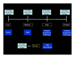
Sometimes the 40 second scan is necessary. There are trade offs for quality, and dose. As an example, if a lesion or cyst is the area of interest to be scanned, then small voxels and more sampling is necessary. Otherwise the radiologist or dentist may not be able to understand the lesion or cyst properties, and therefore not able to diagnosis the patients problem. So a lower resolution scan would be worse since it has little or no values and a second scan would be needed, therefore exposing the patient twice.
The rule is to measure the risk (radiation) with the clinical question being asked, and make the proper choice for scan protocols. As an example, we do NOT need to do a 13cm scan and 20 -40 seconds for an open TMJ or w/Appl. view. Please see attached outline as a sample chart for choosing the best protocol to benefit both the patient and the Dr.

2 Comments:
So according to the chart, EVERY implant scan you do is at .2mm or .25mm @ 40 sec. or is it just the ones that you suspect has some pathology?
Yes, we do high-resolution scans for every implant case (I know Amnon also uses this technique) . If the goal is to see anatomical landmarks every time (like the alveolar canal), then we must choose the correct protocol to achieve those goals. We need to use the tools of imaging to answer the clinical question, and this requires specific imaging solutions. Also, CBCT does not always answer all of the clinical questions; we sometimes need to supplement the scan with other imaging tools like photography and periapicles.
Post a Comment
<< Home