Friday, December 30, 2005

Not enough memory!!!

At the encouragement of Dr. Hatcher, I've recently started taking my scans at .2 & .3 mm. However, I'm finding that when I open the reconstructed data, I get a "not enough memory" message and have to close the program and reopen it to be able to open the data. Is this common or do I have something hogging memory in my computer?

Wednesday, December 14, 2005

Less than 2 seconds


I think we, as radiographers should always consider the ALARA rule when using ionizing radiation: (as low as reasonably achievable), and think about this rule for each and every case we do.

Arun had this comment about the low dose scan:

“We do intend to provide a "child setting" for both normal 20-second scans and 10-second scans, with reduced kVp, mA, or both, optimal combination of which would have to be determined. We hope to get this project going soon. Arun”
This is good news from ISI because we should be able to reduce dose even further. The images I displayed should be better on a small child due to less scatter and smaller head volume. So we should expect the quality to improve a little since this scan was taken on an adult. Here are the attached TMJ images.

Tuesday, December 13, 2005

Less Than 2 seconds


DDI has been testing a protocol to reduce dose to children for orthodontic applications, and we have asked Arun a few questions about this to understand the capabilities of the I-CAT.

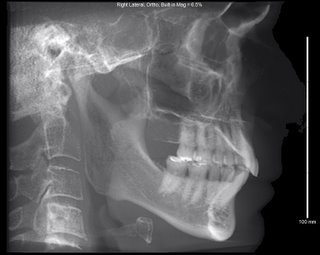
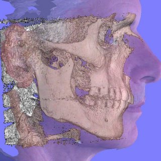
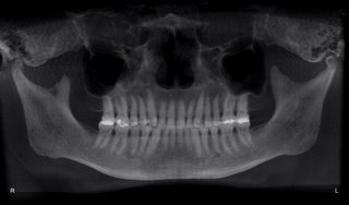
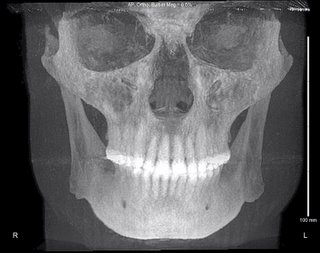
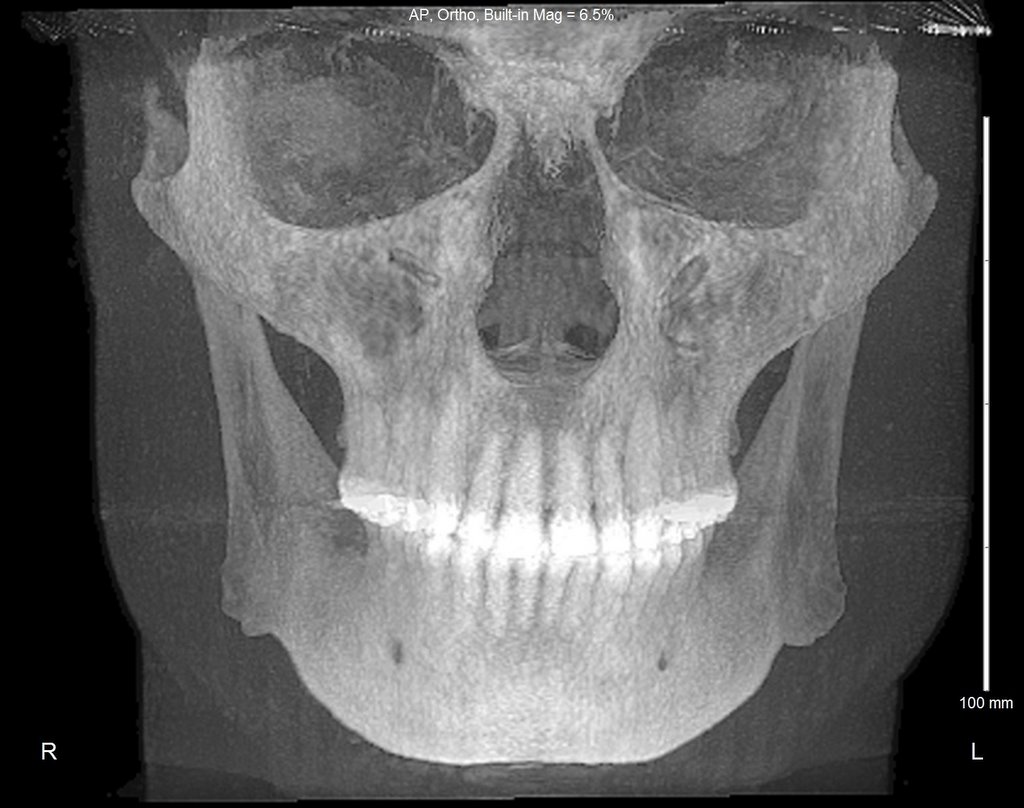
We tested this using the 13cm height with a 10 second scan mode. Then we imported the data into Dolphin 3D and created the typical ortho views plus added views like TM joint, airway and 3D renderings. When I asked Arun how much time the patient was being exposed to the pulsed 10 second scan he said “Less than 1.75 seconds” Arun further stated that “Dose could be lowered further, perhaps dramatically, we (ISI) intend to launch a project to investigate this soon”.

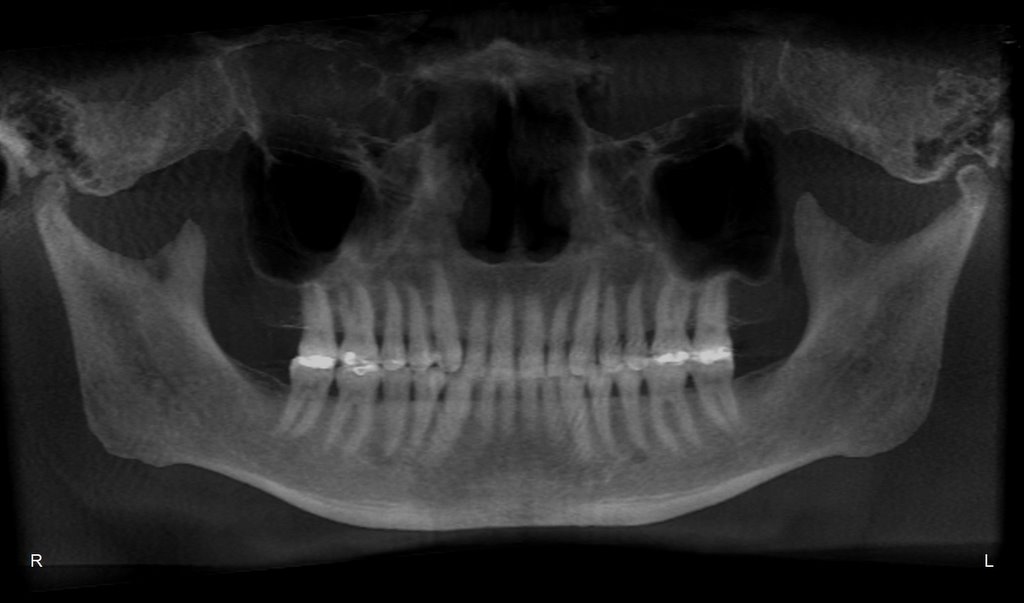
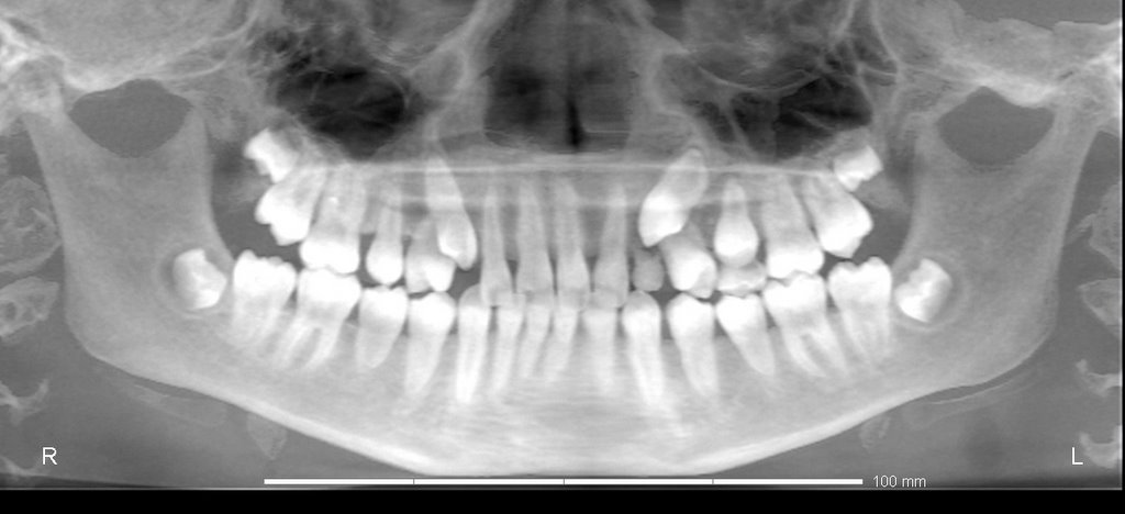
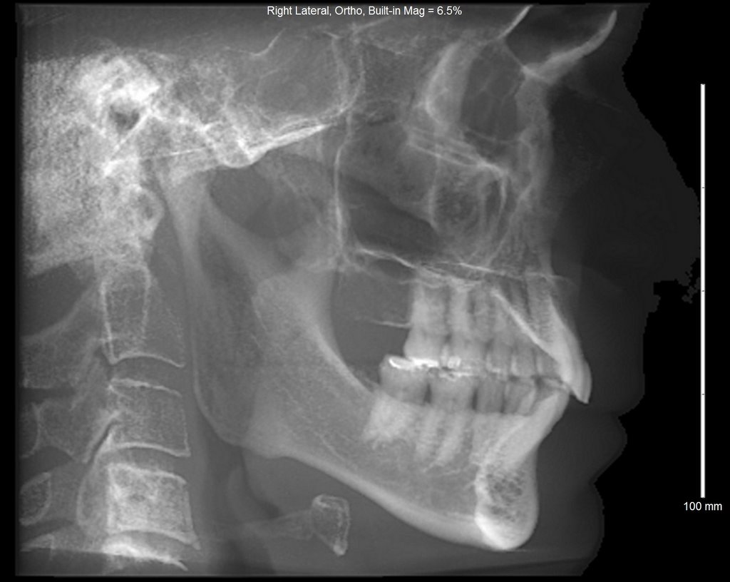
We tested this theory by doing a scan on a orthodontist that was asking the question about mid treatment scans. His thoughts were that we may do a higher resolution scan in the beginning ortho to rule out any abnormalities, and if the case was a “standard” case, then at mid-treatment, we could consider doing a low resolution scan.

The images attached is of the Orthodontist, and the images should be of better quality if we use a child.

Monday, December 12, 2005

MIP Panos


eric....here are the panos....what do you think?

We recently did a MIP pano for maxillary implants sites. Patient had some fluid, mucoceles, or some pathology in sinus (which of course we see pretty often). In this case the patient had very little bone - literally a few mm in some places. When we did a 14mm MIP the fluid (or whatever) in the sinus appears as bone. So, the doc calls and says "what is this? why do I have bone in this pano and not in the other?" Our standard answer is always to use the cross sectional views as the gospel - not pano views. But it was odd. Any comments? What exactly are we seeing in the MIP view?

Also, for the pano view for maxillary implants - we are only mapping the maxilla (duh...). We used to map all the way back to the TMJ just to get the traditional "pano" look but it causes more problems because the mandible is so distorted - or we split the difference and the maxilla is not as accurate. Am I a little slow to figure this out?

Wednesday, December 07, 2005

Updated Dolphin 3D


Scroll down to Dolphin Subsection on this Blog for more infomation to get the newest update for Dolphin 3D...

"AWESOME!" We just received the new Dolphin 3D update. The Dolphin 3D cephalometric profile was already good and now Dolphin has made it even better! It is truly a stunning cephalometric image. Commments on the new panoramic image? Being able to vary the focal trough for the anteriors now allows us to generate a truly pulchritudinous panoramic image worthy of the scrutiny of orthodontists. We now possess the tools (Dolphin 3D and i-CAT) to eliminate the traditional panoramic film & cephalometric profile by utilizing the i-CAT scan. Your orthodontists will have the options of obtaining the TMJ views (lateral & coronal), SMV, cross sectional images. PA skull & more.

Ask your orthodontists if they would like to place Temporary Anchorage Devices (TADs) or mini-implants. We are all familiar with the resistance of children regarding the use of headgear, yet have you considered the adult patient who would be required to wear headgear too? TADs will enable your orthodontist to capture the adult market that refused to wear headgear=more patients. Some orthodontists are not comfortable placing TADs, let them know the scan can be given to the oral surgeon who will place the TADs for them. Explain in detail how you are now able to generate many different images from just one scan and how the orthodontist can see the cross sectional images in the exact area of the TAD site.